Explore Core Facilities

Core facilities play an integral role in our research infrastructure, empowering researchers to break new ground by offering specialized laboratories with state-of-the-art equipment, unique instrumentation and shared services managed by scientists with the technical expertise and experience to help others.

Filter

Category
Location

Alphabet Navigation

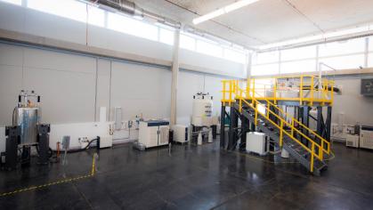
  • High Field NMR Website Image

    The High Field NMR facility provides access to state-of-the art NMR spectroscopy. This includes access to five high field NMR spectrometers, modern high-end solution and solids probes, pulse sequences, sample preparation strategies, and experimental know how, to perform the most up-to-date NMR experiments. The facility is capable of solution and solid-state biomolecular NMR, materials and polymer characterization, and pharmaceutical investigations. The facility provides access and training on the NMR instruments and varying degrees of staff support for novice through experienced users.

    Key Resources
    • Bio-Solution: Available at 800, 700, and 600 MHz, potential experiments include protein dynamics and structure elucidation and protein-ligand binding.
    • Bio-Solids: On the 800 and 600 MHz spectrometers, fast magic-angle-spinning (MAS) probes allow for solution-like investigations of membrane proteins, aggregates, and large complexes.
    • Materials: In addition to the 800 and 600 MHz NMRs, a dedicated 400 MHz instrument is available for work on solid materials such as concrete, polymers, catalysts, and batteries.
    • Service: Various levels of support are available, ranging from access only, to complete data acquisition and analysis.
    Location New Brunswick/Piscataway
    Contact Name Jean Baum
    Contact Email baum@chem.rutgers.edu
  • The imaging core facility offers a broad range of instruments to meet microscopy needs: from bright field and true color imaging, to epifluoresence, laser confocal, super-resolution, multispectral, and live imaging. The facility also offers image analysis software for use, including deconvolution, 3D-reconstruction, and advanced segmentation analysis.

    Request Services

    Key Resources Leica TCS SP8 τ-STED 3X FALCON, Leica Confocal TCS SP8 with Lightning, Nikon Eclipse E800 Bright Field, Zeiss Axiovert 200M, Zeiss LSM510 META (Inverted), Zeiss LSM800 with Airyscan point scanning confocal microscope, Zeiss LSM880 with Airyscan FAST laser scanning confocal, Zeiss Observer Z1 advanced inverted fluorescence microscope
    Location New Brunswick/Piscataway
    Contact Name Karen Schindler
    Contact Email ks804@hginj.rutgers.edu